A Pancoast tumour, also called a superior sulcus tumour, is a lung cancer in the top corner of the lung that grows into the ribs, the nerves and the blood vessels at the base of the neck. It is usually treated as a shoulder or neck problem for months before the cause is found. Treated with chemotherapy and radiotherapy first and then an operation, about half of patients are alive at five years. Dr Lawrence Okiror, Consultant Thoracic and Robotic Surgeon (GMC 6150382), assesses patients at London Bridge Hospital and The Lister Hospital Chelsea within 2–3 days and performs the operation at London Bridge Hospital. Self-referrals welcome.
Last reviewed: October 2026 · Dr Lawrence Okiror FRCS(CTh) FRCSEd(CTh) · GMC 6150382
Pain in the shoulder, often spreading down the inside of the arm to the little and ring fingers, with tingling or a weak grip. A cough or breathlessness is unusual, because the tumour grows outwards into the neck rather than into the airways.
About five weeks of daily radiotherapy with chemotherapy, then a restaging scan, then the operation three to five weeks later. This sequence is the standard because it doubles the chance that the tumour comes out completely.
The top of the lung is removed together with the involved ribs and whatever the tumour has grown into at the base of the neck, through an incision at the front or at the back depending on where the tumour sits. Dr Okiror performs both approaches.
Families write that the tumour was called inoperable because it involves the ribs, the nerves or the blood vessels at the base of the neck. Those are the structures this operation is designed to remove. Involvement of the first rib, the lowest nerve root to the arm or the artery under the collarbone describes the size of the operation. It does not decide whether the cancer can be treated to cure.
What does rule the operation out is spread to the brain or to other organs, a lymph node on the opposite side of the chest, extensive involvement of the nerves above the first thoracic root, or a patient who is not fit for the treatment. Each of those is checked with a specific test, and most are set out below. Where the decision was made by a team that does not perform this operation, a second surgical opinion is a reasonable step. The other reasons people are given are on the told surgery is not an option page.
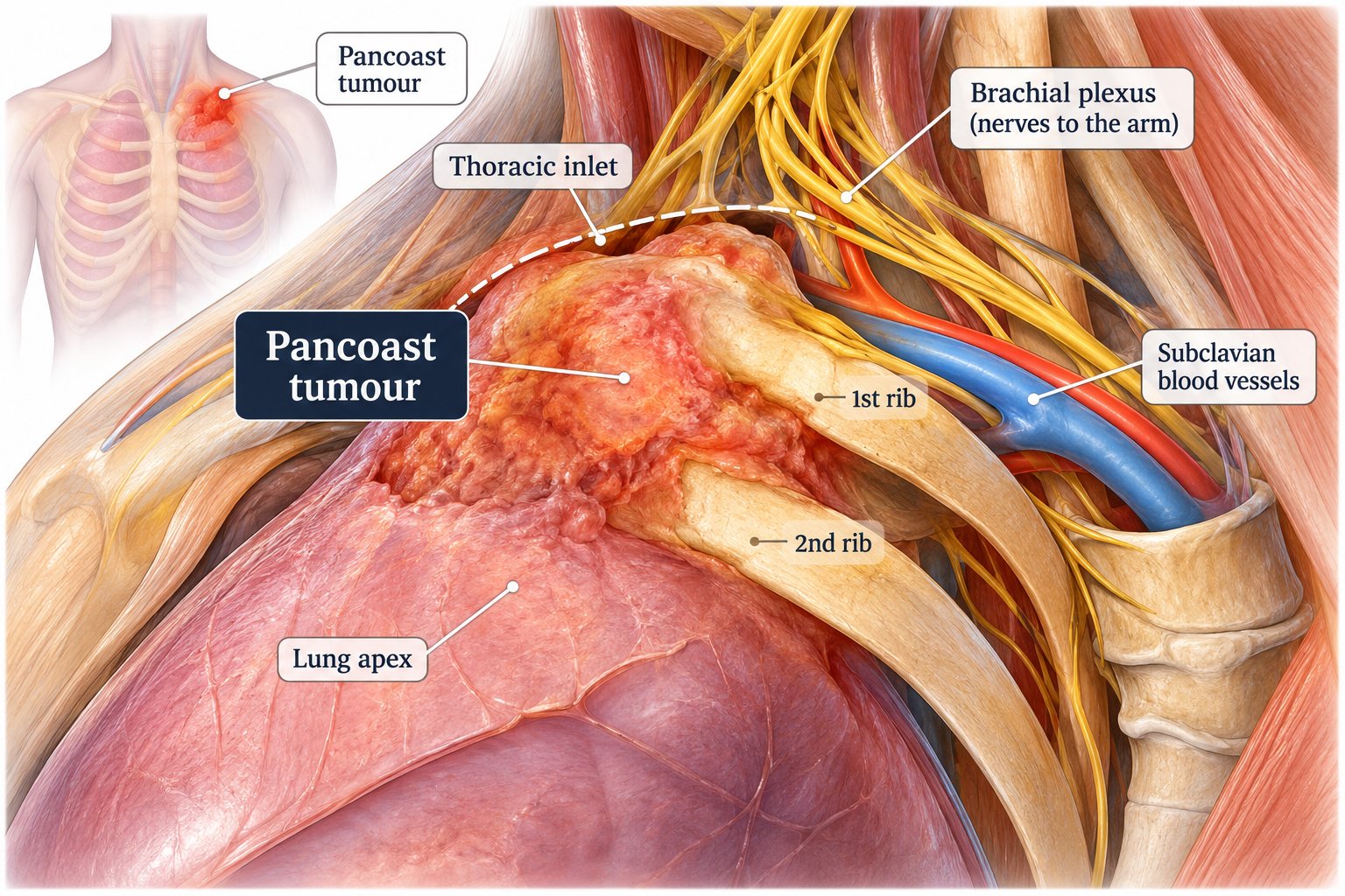
A Pancoast tumour is a lung cancer that starts in the very top of the lung, in the narrow space where the lung meets the base of the neck. That space, the thoracic inlet, is crowded. The first and second ribs form its floor, the nerves to the arm pass over the first rib, and the artery and vein to the arm run beside them. A tumour growing there reaches those structures early, while it is still small, which is why the symptoms are in the shoulder and the arm rather than in the chest.
The nerves the tumour reaches first are the lowest two of the brachial plexus, called C8 and T1. They supply the inside of the forearm, the little and ring fingers, and the small muscles of the hand. Pressure on them causes pain down the inside of the arm, pins and needles in the two outer fingers, and a weak or clumsy grip. Behind those nerves lies a chain of nerves to the eye and the face, and pressure there causes Horner’s syndrome, described below. Pancoast tumours are nearly always non-small cell lung cancers and make up about 3 to 5 per cent of lung cancers [2].
The name comes from Henry Pancoast, the American radiologist who described the pattern in 1924 and 1932. Surgeons use the term superior sulcus tumour for the same cancer. On a scan report it may appear as an apical mass or a tumour at the lung apex.
Because shoulder and arm pain almost always has a shoulder or neck cause, and those causes are investigated first. A 2026 review of 31 studies and 1,328 patients found shoulder pain to be the first symptom in 60 per cent and arm pain in 26 per cent; the commonest wrong diagnoses were a trapped nerve in the neck and arthritis of the shoulder, and the reported average delay to the correct diagnosis ranged from under three months to over a year [1].
The pattern families describe is consistent. An MRI of the spine is normal. A shoulder surgeon arranges an X-ray, an MRI and a steroid injection, and nothing changes. Physiotherapy is tried. The pain is severe, constant, and keeps the patient awake, which is unusual for a shoulder or neck problem. By the time a scan of the chest is requested, the tumour has often grown into the ribs.
The features that point to the chest rather than the shoulder. Pain that is constant rather than related to movement of the shoulder. Pain that runs down the inside of the arm to the little and ring fingers rather than down the outside. Tingling or weakness in the hand. Weight loss. A drooping eyelid on the same side. A history of smoking. Any one of these in a patient whose shoulder and neck treatment has not helped is a reason to request a CT scan of the chest.
The same symptoms, without the weight loss and the eyelid, are also the symptoms of thoracic outlet syndrome, in which the same nerves are compressed between the collarbone and the first rib by a cervical rib or a tight muscle rather than by a tumour. The two conditions sit in the same space and are told apart by the scan, which is one reason a CT of the chest belongs early in the assessment of persistent shoulder, arm and hand pain.
Often it will not. On a plain X-ray the top of the lung sits behind the collarbone and the first rib, and an early Pancoast tumour is hidden there or appears as a small area of thickening at the apex that is easy to pass over. A normal chest X-ray does not exclude the diagnosis. Each of the tests below answers a different question, and all of them are needed before treatment is planned.
The test that finds the tumour. It shows its size, which ribs it has reached, and whether the bone is eroded. It is the scan to ask for when shoulder and arm pain has not settled with treatment of the neck or shoulder.
The test that plans the operation. It shows the nerves to the arm, the artery and vein under the collarbone, and the bones of the spine, and whether the tumour has grown into any of them. NICE recommends an MRI for every suspected tumour at the top of the lung [9]. Which nerve roots are involved decides whether the operation can be done and from which side it is approached.
The tests that set the stage, and they belong before treatment starts rather than after it. The PET-CT checks the lymph nodes and the rest of the body. The MRI of the brain checks for spread there, because the brain is the commonest place a Pancoast tumour recurs after treatment [5]. A finding in another organ on the PET scan, such as a spot on the adrenal gland, should be biopsied where possible before the stage is decided, because the difference between stage 3 and stage 4 decides whether treatment is given with the intention of cure. The reasoning is set out on the stage 4 with one site of spread page.
A needle biopsy through the skin, guided by CT, confirms the cancer type and provides tissue for the molecular tests. It also settles the rarer possibility that a mass at the apex is not a lung cancer at all: a nerve sheath tumour growing from the brachial plexus or an intercostal nerve can occupy the same space, and is treated differently. Lymph nodes in the centre of the chest that light up on the PET scan are sampled by EBUS or mediastinoscopy, and a node above the collarbone is sampled with a needle. Nodes in the centre of the chest, once proven, change the plan, so they are not left to the scan.
Lung function tests and an assessment of general fitness complete the work-up, because the treatment is a course of chemotherapy and radiotherapy followed by a major open operation, and the patient has to be able to tolerate both. How fitness is assessed is set out on the lung function and fitness for lung surgery pages, the full set of tests on the tests and staging page, and what each scan can and cannot show on the chest imaging page.
Horner’s syndrome is a drooping upper eyelid, a smaller pupil and reduced sweating on one side of the face. It happens when the tumour reaches the chain of nerves that runs up the side of the spine to the eye, just behind the nerves to the arm. It is present in about one in six patients at diagnosis in the 2026 review [1], with older series reporting between 14 and 50 per cent [2].
Families notice it before doctors do. One eye looks smaller, the lid sits lower, one side of the face stays dry in the heat. In a patient with unexplained shoulder and arm pain it is the single finding that points most directly to a tumour at the top of the lung, and it is a reason for a CT of the chest the same week.
Horner’s syndrome does not by itself make the tumour inoperable. The nerve chain it affects is removed with the tumour, and the eyelid changes often persist after treatment. Some families report the eyelid lifting as the tumour shrinks during chemotherapy and radiotherapy. Horner’s syndrome is distinct from a bulging eye, which has other causes and is not a feature of a Pancoast tumour.
Pain is the dominant symptom. It is usually severe, it is constant, and it spreads from the shoulder down the inside of the arm to the elbow and the little and ring fingers. Patients describe not being able to sleep, and pain in the shoulder blade and the armpit on the same side. It is the one symptom almost every patient has.
Tingling, numbness and weakness in the hand follow as the tumour presses on the lowest nerve roots. The small muscles of the hand waste, the grip weakens, and fine movements such as buttoning a shirt become clumsy. Arm pain was reported in about a quarter of patients at diagnosis [1]. Swelling of the arm points to the vein under the collarbone being compressed.
Weight loss is common by the time the diagnosis is made, because the pain has been present for months and because the cancer itself causes it. It is also the symptom that most often separates a tumour from a shoulder problem, and it is a reason to look harder.
A cough, coughing up blood and breathlessness are unusual at presentation, because the tumour grows outwards into the neck rather than into the airways. Their absence is one of the reasons the chest is not considered.
Because the operation works better after them. The tumour sits against nerve, bone and vessel, where the surgeon cannot take a wide margin of normal tissue. Giving chemotherapy with radiotherapy first shrinks the tumour and sterilises its edge, so that a complete removal becomes possible more often. For stage 3 lung cancer in general the sequence is now chemotherapy with immunotherapy, as set out on the stage 3 lung cancer page. For a Pancoast tumour the sequence is different, and this section explains why.
In the North American trial that set the standard, 110 patients received two cycles of chemotherapy with five weeks of radiotherapy and then surgery. Four in five went on to the operation. The tumour was removed completely in 94 per cent of those operated on, more than half had no cancer or only minimal cancer left in the specimen, and five-year survival was 44 per cent overall and 54 per cent after complete removal [3]. Before this sequence, five-year survival with surgery alone was around 30 per cent.
The Japanese trial of the same sequence, in 76 patients, reported the operation in 76 per cent, complete removal in 89 per cent of those operated on, and five-year survival of 56 per cent, rising to 70 per cent after complete removal [4]. A later North American trial added further chemotherapy after the operation and found that most relapses were in distant organs, with the brain the commonest site [5], which is why the brain MRI before treatment, and the systemic treatment afterwards, are part of the plan.
The surgical series tell the same story. The Paris unit that developed the anterior approach reported 73 patients operated on through it over 22 years: the tumour was removed completely in 90 per cent, the operative mortality was 2.7 per cent, and 54 per cent were alive at five years and 49 per cent at ten [6]. A 2023 series of 155 patients from Memorial Sloan Kettering reported complete removal in 85 to 94 per cent and a major or complete response to the treatment in the specimen in 55 per cent [7]. A pooled analysis of fifteen studies found that two things decide survival above all others: whether the tumour was removed completely, and whether the lymph nodes were involved [13].
On that evidence, the European guideline, the American guideline and the surgical consensus all recommend chemotherapy with radiotherapy and then surgery for a Pancoast tumour that has not spread to the lymph nodes in the centre of the chest, decided at a multidisciplinary meeting [8, 9, 10]. The operation follows the restaging scan, usually three to five weeks after the radiotherapy ends. One in five patients who start the treatment do not reach the operation, most often because the cancer has spread in the meantime or because they are not well enough, and that is explained at the start rather than discovered at the end.
Sometimes, and the decision is made at the multidisciplinary meeting case by case. For most stage 3 lung cancers, chemotherapy with immunotherapy before the operation is now the standard, and the trials behind it are on the stage 3 page. Those trials did not report on Pancoast tumours as a group, and one of them excluded any patient who was due to have radiotherapy before surgery, which is the standard Pancoast pathway. The guidelines therefore still recommend chemotherapy with radiotherapy for this tumour [8, 10].
The published experience of chemotherapy and immunotherapy without radiotherapy in Pancoast tumours is small. The Paris unit reported six patients treated this way in 2024 and 2025: all six had the tumour removed completely with no cancer left in the specimen, and in three the chest wall could be spared, but the follow-up was six months and the authors asked for a trial before the standard changes [11]. A Dutch trial added immunotherapy to the chemotherapy and radiotherapy rather than replacing the radiotherapy, with a high rate of complete response in the specimen and a high rate of serious side effects [12]. A Spanish trial of chemotherapy with immunotherapy and no radiotherapy, and a Japanese trial of chemoradiotherapy with immunotherapy, are both running and neither has reported [14].
The surgeon’s view. Radiotherapy to the top of the lung leaves scarring in the base of the neck, around the artery, the vein and the nerves to the arm, which is exactly where this operation is difficult and where the risk to the arm lies. Immunotherapy leaves its scarring mainly around the airway and the lymph nodes in the centre of the chest, which is where a lobectomy is difficult but not where a Pancoast resection is. That is why surgeons who do this operation lean towards chemotherapy with immunotherapy where the multidisciplinary meeting agrees. The difference has not been measured in a trial, and Dr Okiror states it as a surgical judgement rather than as evidence.
What this means for a patient is that the sequence recommended will depend on the tumour, on the lymph nodes, on the molecular tests and on the view of the multidisciplinary meeting at the time. Either sequence ends in the same operation, performed after a restaging scan. What happens between the end of treatment and the operation is set out on the surgery after chemotherapy and immunotherapy page, and the trial evidence behind each sequence is reviewed for clinicians in the 2026 clinical reference on lung cancer surgery.
For patients who cannot have the operation, because of spread, because the tumour cannot be removed, or because they are not fit for surgery, chemotherapy with radiotherapy followed by a year of immunotherapy is the treatment, and it is given with the intention of cure. Patients write of thirty radiotherapy sessions, six chemotherapy treatments and a year of immunotherapy, and of the tumour no longer being visible at the end of it. That is this pathway.
For a patient who is fit and whose tumour can be removed, the standard remains chemotherapy, radiotherapy and then the operation. The operation removes the part of the tumour that radiotherapy is least able to clear, where it has grown into bone, nerve and vessel, and it removes the source of the pain. Focused radiotherapy in a few sessions, marketed under several names, is a treatment for small tumours inside the lung. It is not the standard treatment for a tumour that has grown into the ribs and the nerves at the base of the neck.
A patient who has completed chemoradiotherapy without an operation and whose tumour remains or returns at the top of the lung may still be a candidate for surgery in selected cases. That situation is set out on the surgery after radiotherapy page.
Often it does not. In the staging system a lymph node above the collarbone is counted as N3, which for most lung cancers places the disease beyond surgery. A Pancoast tumour is the recognised exception. The node sits directly above the tumour, within the field the operation removes, and many surgical units treat a same-side supraclavicular node as part of the local disease rather than as distant spread [2, 15]. Dr Okiror’s team takes the same view, and the question has been discussed at surgical meetings several times without a settled answer.
The data are limited and should be read as such. A small Paris series of 36 patients found no difference in survival between those with and without nodal involvement, including supraclavicular nodes [16]. The larger Paris series found that 10 per cent of patients had a supraclavicular node in the final specimen and that nodal involvement of any kind was associated with worse survival [6]. Surgery with a same-side node is therefore offered to selected patients after the node has been sampled, after the rest of the staging is clear, and after multidisciplinary discussion, with the understanding that the outlook is less good than with no nodes involved.
A node on the opposite side of the neck or chest is different: it places the disease beyond surgery [15]. Lymph nodes in the centre of the chest, between the lungs, are the stronger adverse factor in every series, and they are sampled before the plan is made. How stage 3 is decided from the lymph nodes, and why nodes seen only on a scan are biopsied, is set out on the stage 3 lung cancer page.
No. A Pancoast tumour growing into the first and second ribs is a lung cancer that has extended directly into the chest wall next to it. In the staging system that is stage 3, not stage 4. It is different from cancer that has travelled through the bloodstream to a bone elsewhere, which is stage 4. The distinction decides the treatment: a tumour that has grown into the ribs beside it is removed with those ribs, as one piece, with the intention of cure.
The involved ribs come out attached to the lung, with an uninvolved margin, and the gap is reconstructed where its size and position require it. The principles are the same as for any lung cancer involving the chest wall and are explained in full on the lung cancer invading the chest wall and ribs page. The difference at the top of the lung is that the ribs are short, curved and close to the nerves and vessels, and that the defect sits under the shoulder girdle, which usually supports it without a rigid reconstruction.
The operation removes the top of the lung, usually as a lobectomy of the upper lobe, together with the first, second and third ribs as the tumour involves them, and whatever else at the base of the neck the tumour has grown into: a nerve root, part of the artery under the collarbone, or part of the spine. It comes out as one piece. Which side it is approached from depends on where the tumour sits, and Dr Okiror performs both approaches.
For tumours sitting against the artery, the vein and the nerves at the front of the inlet. The incision runs along the collarbone and down the front of the chest. It gives the surgeon direct sight of the vessels and the nerve roots before anything is divided, which is what makes it possible to remove and reconstruct a segment of the artery and to protect the nerves. The approach was developed in Paris by Professor Philippe Dartevelle and his unit, who reported the first series in 1993 and the 22-year experience in 2024 [6, 17]. Dr Okiror assisted at operations performed by Professor Dartevelle during his training.
For tumours sitting against the spine and the back of the ribs. The incision curves from the base of the neck around the shoulder blade, and the shoulder blade is lifted to reach the ribs beneath it. It is the original Pancoast operation, described in 1961 [18], and it remains the right approach for a tumour at the back of the inlet. The involved ribs are divided at the spine and at the front, and the specimen is removed with the upper lobe.
The first, second and third ribs as involved. The lowest nerve root to the arm where the tumour has grown into it. The nerve chain to the eye. A segment of the artery under the collarbone, which is then reconstructed, where the tumour involves it [6]. Part of a vertebra where the tumour reaches the spine, in a combined operation with a spinal surgeon. The lung itself, usually as an upper lobectomy, with the lymph nodes in the centre of the chest removed for staging.
This is an open operation. Robotic and keyhole access suit most lung cancer resections, and the reasons are set out on the robotic lung surgery page; a Pancoast resection is done openly because the ribs, the nerves and the vessels have to be handled under direct vision and removed in continuity with the lung. The reconstruction of the chest wall, where it is needed, follows the principles on the chest wall page. In the published series cited on this page the risk of dying from the operation is under 3 per cent [3, 6, 17].
The operation is performed at London Bridge Hospital. The chemotherapy and radiotherapy before it are delivered by the oncology team through the multidisciplinary meeting at London Bridge Hospital, and patients who have had that treatment on the NHS can be transferred for the operation. Dr Okiror consults at London Bridge Hospital and at The Lister Hospital Chelsea, and both are available for the first assessment and for follow-up.
The nerve roots are preserved wherever possible. The lowest root, T1, is taken when the tumour has grown into it. Losing T1 weakens the small muscles of the hand, and most patients tolerate it and adapt. The root above it, C8, is not usually taken, because losing it causes a disabling weakness of the hand [15]. Where the MRI shows the tumour involving C8 or higher, that weighs heavily against the operation at the multidisciplinary meeting, and Dr Okiror says so at the first consultation rather than at the end of the treatment.
The nerves between the removed ribs are taken with them, which leaves an area of altered sensation on the chest wall and under the arm. The nerve chain to the eye is removed with the tumour, so a Horner’s syndrome that was present before the operation persists, and one that was not present may appear. Shoulder movement returns with physiotherapy; the shoulder blade is lifted during the posterior approach and the muscles around it need time to recover.
Where the artery under the collarbone has been removed and reconstructed, the blood supply to the arm is restored at the operation and the arm recovers its function. Patients with a reconstructed artery are followed with scans of the graft.
For most patients the pain eases during the chemotherapy and radiotherapy, as the tumour shrinks away from the nerves, and sleep returns. The operation then removes the tumour from the nerves and the ribs it has been pressing on. Pain is the symptom patients have lived with longest, and relief of it is one of the two aims of treatment alongside cure.
Pain after the operation is planned for before it. A Pancoast resection is a large open operation through the chest wall, and breathing well afterwards depends on being comfortable enough to do it. A paravertebral catheter placed at the operation delivers continuous local anaesthetic for several days, a patient-controlled pump covers the first days alongside it, and the plan is set before the operation, as set out on the pain relief after thoracic surgery page. The chest wall wound and the divided intercostal nerves give most of the early pain, and it settles over weeks.
Pain that persists months after treatment has more than one possible cause: damage to the nerves to the arm by the tumour before it was treated, scarring of those nerves from the radiotherapy, or the chest wall itself. It is assessed rather than assumed, because each cause is treated differently, and nerve pain of this kind responds to specific treatment from a pain specialist. Patients should expect that question to be asked at follow-up, and should raise it if it is not.
The hospital stay is longer than for a keyhole lobectomy, and the return to full activity takes weeks to a few months. Physiotherapy for the shoulder and for breathing starts on the first day. Patients go home when they are walking, eating, and managing their pain with tablets, and the general guidance on the weeks that follow is on the recovery after lung surgery page.
The specimen goes to the pathologist, who reports how much cancer remained after the chemotherapy and radiotherapy, whether the margins are clear, and whether the lymph nodes were involved. Those three findings set the outlook and decide what follows. Further treatment after the operation is given by the oncologist and depends on the pathology and on the molecular tests; for many patients it is a period of immunotherapy. Follow-up is with CT scans at intervals for five years.
Patients who have had the chemotherapy and radiotherapy under an NHS team can be transferred for the operation, and the HCA Medical Concierge team arranges the bed and the insurer authorisation. Patients who have completed the operation privately can return to their NHS oncologist for the treatment that follows, or continue with the oncology team at London Bridge Hospital.
The CT scan, the MRI of the top of the chest if one has been done, the PET-CT, the MRI of the brain, the biopsy result with the molecular tests, and the letter from the multidisciplinary meeting. Tell the practice which hospital did the scans and the images are transferred electronically, usually within 24 to 48 hours. Dr Okiror reviews the images himself before the appointment, and the consultation is spent on the decision.
Situations in which a surgical opinion is reasonable:
Appointments are available within 2–3 days at London Bridge Hospital or The Lister Hospital Chelsea, and by video within 24 hours. Patients outside London, and patients abroad, are seen by video first, and family members can join the same consultation from other cities or countries. A written report follows within 48–72 hours. The consultation fee is £250–£350, and most insurers fund it in full, less any excess written into the policy. Where the operation follows, the hospital issues a written fixed quote after the consultation and before treatment; hospital charges are the largest part of the total, and insurers usually settle the hospital, surgeon and anaesthetist fees in full, less any policy excess. How fees work →
The opinion often confirms the plan already made. What the patient receives in that case is the reason, in writing, with the tests that would change it. Where the plan changes, surgery is usually within a week of completing the investigations, and for a Pancoast tumour the timing is set by the end of the chemotherapy and radiotherapy and the restaging scan. Lung cancer second opinion service →
Questions asked by patients and families after a diagnosis of a Pancoast tumour, and by patients with shoulder and arm pain that has not been explained.
Request a Second Opinion →Or call Jo Mitchelson:
020 7952 2882
Self-referrals welcome. Appointments at London Bridge Hospital and The Lister Hospital Chelsea within 2–3 days, by video within 24 hours. Dr Okiror reviews the scans himself before the appointment and gives a clear answer at the first consultation.
Jo Mitchelson, PA · 020 7952 2882 · pa@lungsurgeon.co.uk
St Thomas’ Hospital #1 UK · Guy’s Hospital #2 UK · London Bridge Hospital #10 UK · Newsweek World’s Best Hospitals 2026