Xiphoid Process Pain
and the Lump at the Bottom of the Breastbone

The hard point many people find at the very bottom of the breastbone is the xiphoid process — a small projection of the sternum that everyone has. It is not a growth and it has not appeared. What usually changes is your ability to feel it: soft tissue over it is lost after weight loss, it hardens with age, or the tissues around it alter after chest or upper abdominal surgery. A xiphoid you can feel but that does not hurt when pressed is normal and needs no treatment. Where firm pressure on it reproduces your exact pain, the diagnosis is xiphoid syndrome — made by examination rather than by scan. Dr Lawrence Okiror, Consultant Thoracic and Robotic Surgeon (GMC 6150382), assesses xiphoid pain at London Bridge Hospital and The Lister Hospital Chelsea within 2–3 days. Self-referrals welcome.

Last reviewed: August 2026 · Dr Lawrence Okiror FRCS(CTh) FRCSEd(CTh) · GMC 6150382

Usually Normal

Everyone has a xiphoid process. Feeling it for the first time after weight loss, after heart surgery, or simply with age is expected. Awareness of a normal structure is not a disease.

Pressure Is the Test

Xiphoid syndrome is diagnosed by examination. Firm pressure over the xiphoid reproduces the pain exactly and it settles when released. No scan makes this diagnosis, and a normal scan does not exclude it.

Treatable When Confirmed

Where the xiphoid is confirmed as the source and symptoms persist, removing it relieves the pain in the great majority. Usually a day case. Selection matters more than technique.

Can you feel it, does it hurt, or both?

Three quite different situations bring people to this page, and they have different answers. You can feel it but it does not hurt. This is much the commonest, and it is normal — the reassurance below is the whole answer. You can feel it and it hurts. Common after heart surgery through the breastbone, and after significant weight loss. It hurts but there is nothing to feel. This is the group who have usually been investigated for months or years.

The single question that separates them is not what the lump looks like, and it is not what a scan shows. It is whether pressing firmly on the tip of the breastbone reproduces the pain you already have. Being able to feel a bone is not a disease. Pain that can be reproduced by pressing on that bone is a diagnosis. Everything on this page follows from that distinction.

  1. The lump is almost always a normal bone you have started to feel.The xiphoid process is present in everyone. Weight loss, ageing, and previous chest or upper abdominal surgery make it easier to feel. Becoming aware of it is not the same as developing a disease.
  2. Its shape varies more than any other part of the breastbone.The xiphoid may point straight down, angle forwards, or be split into two prongs. These variants are normal and present from early life — but an angled or long xiphoid is more likely to be felt, and more likely to catch.
  3. Pressure reproducing your exact pain is what makes the diagnosis.Xiphoid syndrome is diagnosed by examination. Firm pressure over the xiphoid reproduces the pain precisely, and it settles when the pressure is released. Feeling the bone without tenderness is not xiphoid syndrome.
  4. Normal scans do not exclude it.CT, MRI, ultrasound, endoscopy and cardiac tests are performed to exclude other causes. Imaging can measure the angle and length of the xiphoid, but the published series found no relationship between those measurements and outcome after treatment.
  5. Surgery works when the diagnosis is right — and only then.Removal of the xiphoid process is usually a day case and the small published series report substantial pain relief without complications. It does not help a patient whose pain originates elsewhere, which is why selection matters more than technique.

What is the xiphoid process, and where exactly is it?

The breastbone has three parts. The manubrium sits at the top, where the collarbones attach. The body of the sternum lies below it, with the rib cartilages joining along each side. At the very bottom, forming the lowest point of the breastbone, is the xiphoid process — a small projection roughly the size of a thumbnail, sitting immediately above the upper abdomen and directly in front of the top of the stomach.
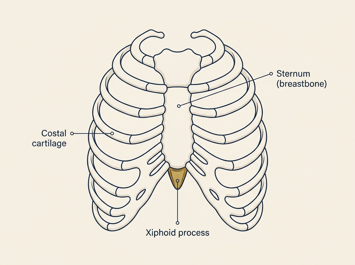
Anatomical illustration of the front of the rib cage, labelling the sternum (breastbone), the costal cartilages joining the ribs to it, and the xiphoid process highlighted at the lower tip of the breastbone
The xiphoid process sits at the lowest point of the breastbone, where the rib cartilages meet in the midline — immediately above the upper abdomen.

It begins life as cartilage and hardens into bone gradually over the course of adult life, a process that is often incomplete until middle age and sometimes never fully complete at all. That slow hardening matters, because it is one of the reasons people become aware of the xiphoid in their forties and fifties having never noticed it before. The structure has not changed shape; it has changed consistency.

Its position explains almost everything patients describe. It sits at the junction between chest and abdomen, where the diaphragm and the upper abdominal muscles attach, immediately in front of the stomach and close to the lower end of the oesophagus. Pain arising here is felt in exactly the region where cardiac pain, reflux and gallbladder pain are also felt — which is why so many people with this problem are investigated thoroughly for all three before anyone examines the bone itself.

Why can I suddenly feel it when I never could before?

This is the question people most want answered, and in almost every case the answer is that the xiphoid has been uncovered rather than altered.

After weight loss

The commonest reason, and an increasingly common one. Fat over the lower sternum is lost and the bone beneath becomes palpable and sometimes visible. This is now frequently noticed after substantial weight loss on a GLP-1 medication such as semaglutide or tirzepatide, and after bariatric surgery. The same experience is reported with collarbones, ribs and the tailbone. Nothing has grown. On GLP-1 medication and chest surgery →

With age

As the xiphoid ossifies it becomes firmer and easier to distinguish from the surrounding tissue. Many people first become aware of theirs in middle age and, finding a hard point that was not obviously there before, assume it must be new. It is the same structure, felt for the first time.

After heart surgery through the breastbone

A sternotomy divides the breastbone and it is rejoined at the end of the operation. The xiphoid sits at the very bottom of that incision, and afterwards it commonly becomes more prominent, more mobile, or tender. Patients describe being unable to lie face down, or to hold anything against the chest. Swelling in the area settles over months in many people; a xiphoid that remains prominent and painful well beyond that is worth examining.

After upper abdominal surgery

Operations in the upper abdomen alter the muscle and fascia that attach around the lower sternum. Pain that begins in this area after gallbladder or other upper abdominal surgery, and that is reproduced by pressing on the xiphoid, deserves examination of the xiphoid specifically rather than further investigation of the original operation.

After a blow, heavy lifting, or a long bout of coughing

A direct blow to the lower sternum can injure the xiphoid, and so can sustained force from heavy lifting or weeks of forceful coughing. This history is easily missed, partly because the injury is often minor at the time and partly because plain X-rays frequently show nothing. Onset that dates from a specific event of this kind is a useful clue.

Or it has always been like that

Some people have a prominent xiphoid from childhood and have simply never thought about it until something drew their attention to it. A prominence that has been unchanged for as long as you can remember, and that does not hurt, is a normal variation.

Why does mine stick out when other people’s do not?

Because the xiphoid is the most variable part of the sternum, and by some margin. In many people it continues straight down in line with the breastbone and is barely detectable. In others it angles forwards, away from the chest, so that the tip sits closer to the skin. In a proportion of people it is split into two prongs — described as bifid. It also varies considerably in length. All of these are normal anatomical variants, set early in life, and none of them is a deformity.

This variability is the reason two people can lose the same amount of weight and have completely different experiences. One feels nothing. The other finds a hard, sharply defined point that seems far too prominent to be normal. The difference is not the weight loss; it is the shape that was always there underneath.

Shape also helps explain the symptoms people describe when the xiphoid does become painful. The published surgical series measure the angle the xiphoid makes with the sternum and its overall length, and in one series of nineteen operated patients the median angle was 149 degrees against a described threshold of 160 — that is, a tip angled distinctly forwards [1]. A forward-angled or elongated xiphoid is more likely to be felt, and more likely to catch against the tissues in front of it when you bend forward, lie flat, or eat a large meal. It is a mechanical description, and it matches what patients report: catching, clicking, and a sensation of something moving rather than something inflamed.

When is a prominent xiphoid completely normal?

A xiphoid process that you can feel, or even see, and that does not hurt when you press on it, is a normal part of your skeleton. It does not need scanning, monitoring, or removing. It is not a tumour. It has not grown — and unless something else changes, it will not.

It is worth saying plainly, because a great many people find this point, cannot identify it, and conclude the worst. A hard lump in the exact midline at the very bottom of the breastbone, unchanged and painless, is the normal anatomy of that region. Equally, not being able to feel yours means nothing either — plenty of people never can, and that is just as normal.

What would make it worth being examined:

When is it xiphoid syndrome?

Xiphoid syndrome — also called xiphodynia — is pain arising from the xiphoid process itself. It was first described in 1955 and it remains uncommon, which is a large part of why it goes unrecognised for so long [1].

The pain sits at the lower end of the breastbone and characteristically spreads — to the upper abdomen, the lower chest, the back between the shoulder blades, the throat, or the shoulders. It is typically worse on bending forwards, on lying flat, and after a large meal, and better sitting upright. People describe it as sharp, or aching, or as a bruise; many describe a catching or clicking sensation on movement or deep breathing.

The defining feature is reproduction. Firm pressure applied directly over the xiphoid brings on the exact pain you already have — not a general tenderness, but your pain — and it settles when the pressure is released. That finding, in someone whose cardiac and upper gastrointestinal investigations are normal, is what makes the diagnosis. The published series describe it the same way: diagnosis rests on physical examination and reproduction of symptoms on manual compression, after other causes have been excluded [1].

This takes seconds to test and it is the step most often skipped — not through carelessness, but because the pain sits precisely where far more common and more dangerous conditions sit, and those are rightly excluded first. The problem is what happens afterwards. Once the heart and the stomach have been cleared, many patients are left without a next step rather than being examined for the chest wall.

Why have all my scans come back normal?

Because this is not a diagnosis that scans make. A normal ECG, a normal echocardiogram, a normal gastroscopy, a normal ultrasound and a normal CT do not exclude xiphoid syndrome, and they were never capable of doing so. They exclude other things — which is a necessary job, and it has to be done first.

Cross-sectional imaging can describe the xiphoid: its angle relative to the sternum, its length, whether it is bifid, whether it has been fractured. That description is worth having. But it does not identify who is in pain, and it does not predict who will be helped by treatment. In the published surgical series, the imaging measurements bore no relationship to how patients did afterwards [1]. The scan describes the shape. The examination finds the source.

In practice, imaging is arranged when there is a specific question to answer — a suspected fracture, a lump that does not behave like a xiphoid, an epigastric hernia, or a cause in the upper abdomen that has not yet been excluded. It is not arranged to confirm a diagnosis that has already been made at the bedside. Bring the results you already have to the consultation: knowing precisely what has been excluded is more useful than repeating it.

What makes it worse — and what should I stop doing?

Stop pressing on it. Repeated prodding, squeezing, deep massage and attempts to manipulate or “pop” the area aggravate exactly what you are testing. It is the commonest reason a tender spot becomes considerably more tender over the weeks before an appointment, and several people work this out for themselves before anyone tells them. Checking whether it still hurts, several times a day, is what keeps it hurting.

Beyond that: the positions that provoke it are worth avoiding while things settle — lying flat, prolonged bending forward, large meals late in the evening, and heavy lifting through the front of the chest. Simple analgesia is reasonable. Alternating ice and heat does no harm but has no particular evidence behind it in this condition.

One thing to be careful about. A great deal is written online about reflux medication in relation to this kind of pain, some of it confidently wrong. If you are taking a proton pump inhibitor or another reflux treatment, do not stop it, start it, or change the dose on the strength of something you have read. That decision belongs with the clinician who prescribed it, and it is worth raising directly with them.

What else could this be?

Not everyone who reaches this page has xiphoid syndrome, and it would be doing you no favours to imply otherwise. Several conditions produce pain in the same small area, and telling them apart is the work of the consultation.

Slipped rib syndrome

The closest and most frequently confused. The problem lies in the lower rib cartilages at the edge of the rib cage, but the pain is very often felt at the bottom of the breastbone, and the clicking and catching sound identical when described. It is distinguished by where the tenderness actually is, and by a specific examination manoeuvre. Slipped rib syndrome →

Costochondritis and Tietze syndrome

Inflammation of the joints between the ribs and the breastbone, generally higher up and often on both sides. Tietze syndrome adds visible swelling. Both can coexist with xiphoid tenderness, which is one reason people are given the label and then find it does not explain everything. Unexplained chest pain →

Reflux and upper abdominal causes

Reflux, gastritis, gallbladder disease and an epigastric hernia all produce pain in this region, and reflux frequently coexists with xiphoid pain rather than replacing it as an explanation. An epigastric hernia in particular can present as a lump near the midline just below the xiphoid and is easily mistaken for it.

A lump that is not the xiphoid

If what you can feel is off the midline, mobile under the skin, or soft rather than hard, it is unlikely to be the xiphoid process. Chest wall lumps have their own set of causes, most of them benign. Chest wall lump →

Cardiac causes come first

No chest wall diagnosis should be settled on until cardiac causes have been properly excluded. If you have not had that assessment, that is the first step, not this page. New, severe, or exertional chest pain needs urgent medical attention rather than an outpatient appointment.

How is xiphoid syndrome treated?

Most people do not need an operation. Avoiding the provoking positions, leaving the area alone, simple analgesia and time settle a good proportion of cases. Where pain persists despite that, and the xiphoid has been confirmed as the source, the xiphoid process can be removed.

The operation is small. The xiphoid is separated from the lower end of the sternum and removed through a short incision over it, under general anaesthetic. It is usually a day case. The xiphoid has no function that is lost by removing it.

What the published results show. The evidence base is small, because the condition is uncommon. In a series of nineteen patients, pain scores fell from a median of 8 out of 10 before surgery to 0 afterwards, seventeen of the nineteen had a clinically meaningful improvement, and there were no post-operative complications; the median operating time was 29 minutes [1]. A second series of nineteen patients, published the same year, reported improvement in 94% and complete freedom from pain in 63% at a mean of 34 months, again with no surgical complications [2].

Those are good results, and they should be read for what they are: two small retrospective series from single centres, in a condition where the entire published surgical experience runs to a few dozen operated patients. There is no randomised trial and there is unlikely ever to be one. The honest summary is that removing the xiphoid reliably relieves pain in patients whose pain is genuinely coming from the xiphoid — and does nothing at all for anyone else.

Which is why the consultation matters more than the operation. The commonest fear among people who have lived with this pain for years is not the surgery — it is another intervention that does not work. Being told clearly that the xiphoid is not the source, and what the alternatives are, is a legitimate and often better outcome of a consultation than being offered an operation.

Which specialist should I actually see?

A thoracic surgeon. The sternum, the xiphoid process, the costal cartilages and the ribs are thoracic anatomy, and chest wall pain is thoracic surgical territory. This is worth stating plainly, because the single thing most people with this problem have in common is that nobody has told them which specialty owns it. Cardiology has excluded the heart. Gastroenterology has excluded the stomach. Physiotherapy and manual therapy have been tried. And then the trail goes cold.

Dr Okiror assesses xiphoid and chest wall pain at London Bridge Hospital and The Lister Hospital Chelsea, with outpatient consultations also available at the HCA clinics in Canary Wharf and the City of London. The first appointment is an examination, a review of what has already been excluded, and a clear statement of whether the xiphoid is the source of the pain. Where it is not, you will be told so, and told what to pursue instead.

Private appointments are typically available within 2–3 working days. Self-referrals welcome. Bring your cardiac and endoscopy reports and any imaging you already have. Second opinions are available for patients already under another team’s care.

Questions About
Xiphoid Process Pain

Questions most commonly asked by people who have found a lump at the bottom of the breastbone, or who have pain there that has not been explained.

Book an Assessment →

Or call Jo Mitchelson:
020 7952 2882

What is the hard lump at the bottom of my breastbone?
In the great majority of people it is the xiphoid process — the small projection at the lower tip of the sternum, present in everyone from birth. It is not a growth and it is not new. What has usually changed is your ability to feel it: soft tissue over it has been lost, or it has hardened with age, or the tissues around it have altered after an operation. A xiphoid process that can be felt but does not hurt when you press on it needs no treatment. If it is tender, growing, or sits to one side of the midline rather than at the centre, it should be examined.
Why can I suddenly feel my xiphoid process after losing weight?
Because the fat that was covering it has gone. The xiphoid has not moved or grown — it has been uncovered. This is now a common reason for people to notice it for the first time, particularly after substantial weight loss on a GLP-1 medication such as semaglutide or tirzepatide, or after bariatric surgery. The same thing happens with collarbones, ribs and the tailbone. Feeling a bone that was previously buried is expected, not a sign that something has gone wrong.
Why does my xiphoid stick out when other people’s do not?
The xiphoid is the most variable part of the sternum. In some people it points straight down in line with the breastbone; in others it angles forwards away from the chest, and in a proportion of people it is split into two prongs — described as bifid. It also varies in length. These are normal anatomical variants present from early life, not deformities. An angled or elongated xiphoid is simply more likely to be felt, and more likely to catch against the tissues in front of it when you bend, lie flat, or after a large meal.
What is xiphoid syndrome?
Xiphoid syndrome, also called xiphodynia, is pain arising from the xiphoid process itself. It was first described in 1955 and remains uncommon. The pain sits at the lower end of the breastbone and often spreads to the upper abdomen, the lower chest, the back, the throat or the shoulders, which is why it is so often mistaken for heart or stomach disease. It is characteristically worse on bending forwards, lying flat, after a large meal, and on direct pressure. The defining feature is that firm pressure on the xiphoid reproduces the patient’s exact pain, and the pain settles when the pressure is released.
Why have all my scans come back normal?
Because xiphoid syndrome is a clinical diagnosis, not a radiological one. Normal cardiac tests, a normal gastroscopy, a normal ultrasound and a normal CT do not exclude it. Scans are done to rule other things out, not to make this diagnosis. A CT or MRI can describe the shape of the xiphoid — its angle relative to the sternum, its length, whether it is bifid — but the published surgical series found no relationship between those measurements and how patients did after treatment. The scan describes the anatomy; the examination identifies the source of the pain.
Should I keep pressing on it to see if it still hurts?
No. Repeatedly pressing, squeezing, massaging or attempting to manipulate the xiphoid tends to aggravate the very thing you are testing, and it keeps the area inflamed and sore. It is also the single most common reason a tender area becomes more tender over the weeks before an appointment. Leave it alone. If you are taking medication for reflux, do not stop or change it on the strength of anything you have read online — that decision belongs with the clinician who prescribed it.
What else could this pain be?
Several conditions produce pain in the same area. Slipped rib syndrome arises from the lower rib cartilages and is often felt as pain at the bottom of the breastbone even though the problem lies to one side. Costochondritis and Tietze syndrome affect the joints between the ribs and the sternum higher up. Reflux, an epigastric hernia, and gallbladder disease all produce upper abdominal and lower chest pain. Cardiac causes must be excluded before any chest wall diagnosis is settled on. Distinguishing between these is the purpose of the consultation, and not everyone who reaches this page will have xiphoid syndrome.
How is xiphoid syndrome treated?
Most people are managed without an operation: avoiding the positions and pressures that provoke the pain, simple analgesia, and time. Where symptoms persist and the xiphoid has been confirmed as the source, the xiphoid process can be removed. The published series are small but consistent — in one series of 19 patients, pain scores fell from a median of 8 to 0, seventeen of the nineteen had a clinically meaningful improvement, and there were no post-operative complications. A second series of 19 patients reported improvement in 94% and complete freedom from pain in 63%, with no surgical complications. It is usually a day case. It is not an operation to undertake unless the diagnosis is secure, because removing the xiphoid does not help a patient whose pain is coming from somewhere else.
Which specialist should I see for xiphoid pain?
A thoracic surgeon. The sternum, the xiphoid process, the costal cartilages and the ribs are thoracic anatomy. Most patients with this problem have already been through cardiology and gastroenterology, and sometimes physiotherapy or chiropractic, without anyone having named the specialty that owns the chest wall. If your cardiac and upper gastrointestinal investigations are normal and the pain is reproduced by pressing on the lower breastbone, a thoracic surgical opinion is the appropriate next step.
Do I need a GP referral?
Self-referrals welcome — no GP letter is needed before booking. Private appointments at London Bridge Hospital and The Lister Hospital Chelsea within 2–3 days. Bring any imaging or test results you already have, including cardiac and endoscopy reports, so that the assessment starts from what has already been excluded. New consultations from £250. Most major insurers accepted. Second opinions also available.
  1. Bakens MJAM, et al. A case series: xiphoidectomy for xiphodynia, a rare thoracic wall disorder. Journal of Thoracic Disease 2021.
  2. Operative results after xiphoidectomy in patients with xiphodynia. Retrospective case series, 19 consecutive patients, 2014–2017. Published 2021.

A five-minute examination
can settle a question you have carried for years.

Self-referrals welcome. Private appointments at London Bridge Hospital and The Lister Hospital Chelsea within 2–3 days. Dr Okiror examines the chest wall personally, reviews what has already been excluded, and tells you clearly whether the xiphoid is the source of your pain — or whether it is not.

Book an Assessment → Second Opinion

Jo Mitchelson, PA  · 020 7952 2882 · pa@lungsurgeon.co.uk

St Thomas’ Hospital #1 UK · Guy’s Hospital #2 UK · London Bridge Hospital #10 UK · Newsweek World’s Best Hospitals 2026

Continue Reading

Slipped Rib SyndromeThe closest differential — lower rib cartilage pain that is often felt at the bottom of the breastbone and frequently attributed to reflux. Unexplained Chest PainChest wall causes of pain after cardiac investigation has come back normal — costochondritis, Tietze, slipped rib, thoracic outlet syndrome. Chest Wall LumpFor a lump that is off the midline, mobile, or soft — lipoma, sebaceous cyst, schwannoma, lung hernia and rib lesions. Slipped Rib Second OpinionIndependent review for patients already given a chest wall diagnosis, or offered surgery, elsewhere. Rib Fracture FixationWhere chest wall pain follows an injury — surgical stabilisation for displaced fractures and persistent post-traumatic pain. Specialist Second OpinionIndependent review of your imaging, investigations and diagnosis before committing to any treatment plan.
📅Book 📞020 7952 2882