← All Conditions

Sleeve Resection & Sleeve Lobectomy
Lung-Sparing Surgery for Central Tumours

A tumour growing in or into the main bronchus does not automatically mean losing the lung. In a sleeve resection the affected segment of airway is cut out and the cut ends are sewn back together, so that the lung beyond it keeps working. Where the tumour is confined to the wall of the bronchus — most often a carcinoid tumour — the airway alone is removed and no lung tissue is lost at all. Where a lobe is involved as well, the lobe and the bronchial segment come out together as a sleeve lobectomy and the remaining lung is reconnected. Dr Okiror performs bronchial sleeve resection with or without lung resection, sleeve lobectomy, and pulmonary artery and double sleeve reconstruction, privately at London Bridge Hospital and The Lister Hospital Chelsea and in his NHS practice at Guy’s and St Thomas’ NHS Foundation Trust, where he leads the Central Airways Service. Self-referrals welcome.

Last reviewed: September 2026 · Dr Lawrence Okiror FRCS(CTh) FRCSEd(CTh) · GMC 6150382

The Airway Is Rebuilt

A circumferential segment of bronchus is removed with the tumour and the cut ends are hand-sewn together. Air passes into the lung beyond it again.

Sometimes No Lung Is Removed

Where the tumour sits within the bronchial wall, the airway alone is resected and reconstructed. The whole lung is preserved.

14,194 Patients

Pooled across 27 studies, survival at one, three and five years was higher after sleeve lobectomy than after pneumonectomy.

What a Sleeve Resection
Actually Is

The airways branch like a tree. The trachea divides into the left and right main bronchi, and each main bronchus divides again into the bronchi supplying each lobe. A tumour that arises at one of those junctions — at the origin of a lobar bronchus, or in the main bronchus itself — sits at the point where the airway to the whole lung passes. The traditional answer to that problem was to remove the entire lung, because removing the lobe alone would leave tumour behind at the cut edge of the bronchus.

A sleeve resection answers it differently. Instead of removing everything beyond the tumour, the surgeon removes a complete ring — a sleeve — of bronchus containing the tumour, with a clear margin above and below it, and then joins the two cut ends of the airway together. The join is a hand-sewn anastomosis. Once it has healed, air passes through it into the lung beyond exactly as it did before.

The first operation of this kind was performed in London in 1947, by Sir Clement Price Thomas at the Brompton Hospital, for a tumour of the bronchus. It has been standard practice for central airway tumours for decades since. It accounts for a small share of lung cancer operations — in the region of 6 to 8 per cent of resections — which is why it is worth asking about specifically rather than assuming it has been considered.

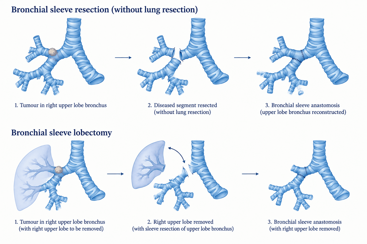
Diagram of bronchial sleeve resection without lung resection and bronchial sleeve lobectomy, each shown in three stages: tumour in the right upper lobe bronchus, resection of the diseased segment, and bronchial sleeve anastomosis
The two operations compared. Above: a bronchial sleeve resection without lung resection — the diseased segment of bronchus is removed and the upper lobe bronchus reconstructed, with the lobe left in place. Below: a bronchial sleeve lobectomy — the right upper lobe is removed together with a sleeve of the adjacent bronchus, and the remaining airway is reconstructed. In both, the third panel shows the completed anastomosis.

The distinction that matters

Patients are frequently told that they need “a sleeve” without being told which of the two operations is meant. They are not the same. One removes a lobe of lung; the other removes no lung at all. Which applies depends on whether the tumour is confined to the wall of the bronchus or has grown outward into the lobe beyond it.

Sleeve Resection Against
Removing the Whole Lung

Four bodies of evidence bear on the same question: where a central tumour can be removed either by taking the whole lung or by resecting and rebuilding the airway, which operation should be done? The comparisons are observational rather than randomised, and that qualification matters — but they point consistently in one direction.

27 Studies · 14,194 Patients

The Largest Pooled Comparison

Translational Lung Cancer Research · 2019 · 4,145 sleeve lobectomy, 10,049 pneumonectomy

Overall survival was significantly higher after sleeve lobectomy at one, three and five years. The pneumonectomy group had higher operative mortality, higher 30-day mortality, and a higher rate of distant recurrence. In patients with N0 and N1 disease, five-year survival after sleeve resection exceeded that after pneumonectomy; in N2 disease the difference at three and five years was not statistically significant.

The nodal subgroup finding is the important detail. Where the mediastinal nodes are involved, the extent of the lung resection is no longer what drives the outcome.

19 Studies · 3,878 Patients

Operative Risk

Shi et al. · World Journal of Surgical Oncology · 2012

Pooled postoperative mortality was 2.91% after sleeve lobectomy against 5.86% after pneumonectomy — an odds ratio of 0.50. The pooled hazard ratio for overall survival was 0.63 in favour of sleeve resection. Rates of postoperative complications and of locoregional recurrence did not differ significantly between the two operations.

Locoregional recurrence is the objection most often raised against sleeve resection — that leaving the lung behind leaves disease behind. The pooled data do not support it.

Extended Sleeve · 431 Patients

When More Than One Lobe Is Involved

Meta-analysis with reconstructed time-to-event data · 2022 · 9 studies

Extended sleeve lobectomy — where more than one lobe is removed and the airway reconstructed — was compared with pneumonectomy using pooled individual patient data. Overall survival favoured the sleeve group (hazard ratio 0.63) as did disease-free survival (hazard ratio 0.57), and both findings held on sensitivity analysis.

This matters because extended sleeve is the operation that most often stands between a patient and a pneumonectomy when the disease is bulkier than a simple sleeve can clear.

Approach · 188 Patients

Robotic, Thoracoscopic and Open

Propensity score-weighted comparison · JTCVS · 2020 · robotic 49, VATS 73, open 66

After weighting, there was no difference in 90-day mortality or morbidity between the three approaches. The robotic group had less blood loss, shorter operative time and shorter drainage time, with no positive bronchial margin and no conversion. Separate series of completely portal robotic airway sleeve resection report a median stay of three days with no 30- or 90-day mortality and no anastomotic stricture at a median of 18 months.

These are small, selected series from experienced centres. They establish that a robotic sleeve resection is a legitimate operation in the right case — not that it is the right operation in every case.

The qualification that belongs with all of this. None of these comparisons is randomised, and patients selected for pneumonectomy tend to have larger, more central and more node-positive tumours than those selected for sleeve resection. Part of the survival difference is a difference in disease rather than a difference in operation. A sleeve resection is chosen because a complete resection can be achieved with a sound reconstruction — not simply because it preserves lung. Where the airway cannot be cleared and rebuilt, removing the whole lung is the correct operation and the evidence above does not argue against it.
Bronchial Sleeve Resection
Without Removing Any Lung

This is the operation most patients do not know exists. Where a tumour grows within the wall of the bronchus and does not extend outward into the surrounding lung, the whole problem is a segment of airway. That segment is removed and the airway rebuilt. The lung beyond it is untouched, re-expands after the operation, and continues to work.

The tumours that behave this way are, most commonly, bronchial carcinoid tumours. These are slow-growing neuroendocrine tumours which often arise in a main or lobar bronchus, frequently in younger patients, and which are commonly mistaken for asthma for months or years because they cause wheeze, cough and repeated infection in the same part of the lung. Typical carcinoid tumours are cured by complete surgical removal in the great majority of cases. Removing an entire lung from a young patient to treat a tumour sitting inside a centimetre of bronchus is a poor trade where a sleeve resection can clear it.

The same principle applies to some rarer airway tumours — low-grade mucoepidermoid carcinoma, and adenoid cystic carcinoma, which spreads along the inside of the airway wall and therefore needs generous margins along its length. These are discussed further on the rare chest tumours page.

Dr Okiror has performed bronchial sleeve resection without lung resection at the left main bronchus and at the bronchus intermedius — the segment of airway between the right upper lobe and the lower part of the right lung, which is the commonest site for a carcinoid tumour that can be treated this way.

A patient with a carcinoid tumour in the bronchus intermedius who is offered a pneumonectomy, and a patient with the same tumour who is offered a bronchial sleeve resection, are being offered two very different futures. The question to ask is whether the tumour is confined to the wall of the airway — and that is answered by bronchoscopy and CT before surgery, and confirmed by inspection at the operation.

Sleeve Lobectomy
The Alternative to Pneumonectomy

Where the tumour involves both the lobar bronchus and the lobe it supplies, the lobe is removed — as in a standard lobectomy — together with a circumferential segment of the adjacent main bronchus. The two remaining cut ends of the airway are then joined, reconnecting the rest of that lung to the trachea. The commonest version is a right upper lobe sleeve lobectomy, where the right upper lobe and a sleeve of the right main bronchus are removed and the bronchus intermedius is sewn to the main bronchus.

The comparison to hold in mind is what the alternative costs. A right pneumonectomy removes around 55 per cent of the lung tissue and the corresponding share of the pulmonary vascular bed. A right upper lobe sleeve lobectomy removes the upper lobe and keeps the rest. For a patient with limited breathing reserve, that difference decides whether surgery is possible at all — which is why borderline lung function is one of the strongest indications to attempt a sleeve reconstruction.

A sleeve lobectomy is also indicated where bulky lymph nodes at the origin of a lobar bronchus make a standard lobectomy impossible — the node involvement extends into the bronchial wall, so the bronchus must be resected further back than a lobectomy allows.

How the join is made

Dr Okiror’s technique for the bronchial anastomosis is described in the chapter he co-authored on primary lung tumours in Tips and Tricks in Thoracic Surgery (Springer, 2018): absorbable suture throughout, running continuously along the membranous back wall of the bronchus and placed as interrupted stitches around the cartilaginous front wall. The membranous and cartilaginous parts of the airway behave differently and are sewn differently for that reason.

Arterial Sleeve and
Double Sleeve Resection

The pulmonary artery runs alongside the bronchus, and a central tumour frequently involves both. Involvement of the interlobar pulmonary artery has historically been one of the standard reasons to proceed to pneumonectomy: if the artery to the remaining lobes cannot be preserved, the lobes cannot be preserved either.

The artery can also be reconstructed. Where tumour involves a segment of the pulmonary artery, that segment is resected and the artery repaired or reconstructed — a pulmonary artery sleeve, also called an arterial sleeve. Where the bronchus and the artery are both involved, both are resected and both reconstructed in the same operation. That is a double sleeve, or bronchovascular sleeve resection.

It is a longer and more demanding operation than a bronchial sleeve alone, it requires control of the pulmonary artery above and below the resected segment, and it is performed in a minority of units. Its value is straightforward: it moves the boundary of what can be preserved. Patients whose tumour involves both structures, and who would otherwise be offered a pneumonectomy, may keep the rest of that lung.

Dr Okiror performs pulmonary artery sleeve and double sleeve resection. As with bronchial sleeve resection, the operation is undertaken only where a complete resection can be achieved; where the artery cannot be reconstructed with a clear margin, the correct operation is a pneumonectomy.

How the Airway Is Assessed
During the Operation

In Dr Okiror’s practice a sleeve resection includes bronchoscopy at three separate points, all under the same anaesthetic on the day of surgery. This is how he conducts the operation; it is not a separate procedure or an extra admission.

At the start

Before the resection begins, the airway is inspected from inside to establish how far the tumour extends along the bronchus in both directions. That determines where the bronchus will be divided. A CT scan shows the bulk of a tumour well; it is less reliable about how far disease creeps along the airway wall, and that distance is precisely what sets the resection margin.

After the anastomosis

Once the two ends of the bronchus have been sewn together, the join is inspected from inside. The mucosal edges should meet accurately and the lumen should be open and round. A problem identified at this point can be corrected while the chest is still open, which is not true of a problem identified afterwards.

At the end

Before the patient is woken, blood and secretions are cleared from the airway and the reconstruction is confirmed to be patent. Retained secretions behind a fresh anastomosis are a cause of early trouble, and clearing them before the patient leaves theatre is part of preventing it.

Diagnostic bronchoscopy before admission is a separate matter and is usually already part of the work-up by the time a patient reaches a surgical opinion. Airway assessment and intervention outside the context of resection is covered on the central airway interventions page.

Open or Robotic —
How the Approach Is Chosen

Most of Dr Okiror’s lung cancer operations are performed robotically or by keyhole technique. Sleeve resection is the part of the practice where that is not the default. Open thoracotomy remains his most common approach to a sleeve resection, and it remains the standard for complex airway reconstruction generally.

He does perform sleeve resection robotically in selected cases. What decides the approach is the reconstruction: the extent of airway to be resected, the position of the anastomosis and how accessible it is, the quality of the bronchial tissue to be joined, whether the pulmonary artery is involved as well, and whether previous treatment has scarred the tissue planes. Where all of those favour it, the operation can be completed robotically to the same standard. Where they do not, an open approach gives the control the reconstruction needs.

Patients who have read about minimally invasive surgery sometimes hear an open approach as a step backwards. It is not. The access is chosen to fit the operation, in both directions: a lobectomy is done robotically because that suits it, and a complex airway reconstruction is done openly for the same kind of reason. An anastomosis that leaks or narrows is the complication that matters most in this operation, and nothing is traded against getting it right.

In this practice

Across six audited years of SCTS returns, Dr Okiror performed 14 sleeve lobectomies. Over the two most recent audited years, six patients underwent sleeve resection for every one who underwent pneumonectomy. That ratio reflects the case mix referred and the anatomy encountered in those particular patients; it is not a target, and it is not a claim that a sleeve resection can replace a pneumonectomy whenever one is proposed.

Who a Sleeve Resection
Is and Is Not For

Considered

Non-small cell lung cancer at the origin of a lobar bronchus or involving the adjacent main bronchus · bronchial carcinoid tumours · low-grade salivary-type airway tumours · bulky N1 nodes at a lobar orifice preventing standard lobectomy · patients whose breathing reserve will not tolerate pneumonectomy · patients whose tumour has responded to chemoimmunotherapy and pulled back from the structures it originally involved

Not suitable

Tumour extending along the bronchus beyond what can be resected with a clear margin · involvement of every lobar orifice on that side · extensive longitudinal involvement of the main pulmonary artery that cannot be reconstructed · disease crossing the fissure into more than one lobe in a pattern a sleeve cannot clear · any situation in which the reconstruction would be achieved only by accepting an incomplete resection

Everyone considered for a sleeve resection is assessed as though a pneumonectomy might be necessary. Occasionally the disease at operation proves more extensive than the imaging suggested, and completing the resection means removing the whole lung. Fitness is therefore assessed against the larger operation, with lung function testing and, where reserve is limited, cardiopulmonary exercise testing — set out on the lung function and fitness pages. Consent for a sleeve resection includes that possibility, discussed before the day rather than explained afterwards.

Recovery and
Follow-Up

The early recovery follows the pattern of the approach used. Where the operation has been performed openly, the hospital stay is longer than after a keyhole lobectomy and pain control is planned in advance rather than improvised afterwards — the approaches used are described on the pain relief after thoracic surgery page. General guidance on the weeks afterwards is on the recovery after lung surgery page.

What is specific to a sleeve resection is the anastomosis. It needs to heal, and healing depends on a good blood supply to the bronchial ends and on the lung beyond it inflating properly. Clearing secretions in the first days matters more than after a standard lobectomy, and physiotherapy starts early for that reason.

Follow-up is clinical and by CT. Bronchoscopy afterwards is not routine; it is arranged where something raises a question about the join — a cough that persists, a wheeze localised to one side, breathlessness that does not settle in the expected way, or an infection recurring in the same part of the lung. Narrowing at the anastomosis is uncommon and is treatable when it is found early, which is the reason those symptoms are worth reporting rather than waiting out.

Where the pathology shows nodal involvement, adjuvant treatment is discussed at the multidisciplinary team meeting in the usual way. Having had a sleeve resection rather than a pneumonectomy generally makes systemic treatment easier to tolerate, because more lung has been kept.

The other side of the decision

Sometimes removing the whole lung is the right operation.

Where the airway cannot be cleared and rebuilt, a pneumonectomy achieves what a sleeve resection cannot, and it is well tolerated in properly selected patients. The indications, the fitness assessment, and what life with one lung is actually like are set out separately.

Pneumonectomy →

Questions About
Sleeve Resection

Questions asked by patients who have been told that a tumour involves the main bronchus, and by those weighing a sleeve resection against removal of the whole lung.

Request a Second Opinion →

Or call Jo Mitchelson:
020 7952 2882

Who performs sleeve resection in London?
Dr Lawrence Okiror performs sleeve resection privately at London Bridge Hospital and The Lister Hospital Chelsea, and in his NHS practice at Guy's and St Thomas' NHS Foundation Trust, where he also leads the Central Airways Service. The operations he performs personally are bronchial sleeve resection with or without lung resection — including of the left main bronchus and the bronchus intermedius — sleeve lobectomy, sleeve tracheal resection, pulmonary artery sleeve and double sleeve resection, and pneumonectomy. Carinal resection and sleeve pneumonectomy are undertaken at a specialist airway centre. Across six audited years of SCTS returns he performed 14 sleeve lobectomies. Sleeve resection accounts for a small proportion of lung cancer operations nationally, so the relevant question when choosing a surgeon is not only whether they can perform it but whether they consider it before recommending removal of the whole lung.
Can a sleeve resection be done robotically?
Yes, in selected cases. Dr Okiror performs some sleeve resections robotically, but open thoracotomy remains his most common approach for this operation and is the standard for complex airway reconstruction. The deciding factors are the extent of the resection, the position and quality of the airway to be joined, and whether the reconstruction can be completed to the same standard through a minimally invasive approach. Published comparisons support robotic sleeve resection in appropriate hands: a propensity score-weighted study of 188 patients found no difference in 90-day mortality or morbidity between robotic, thoracoscopic and open groups, with less blood loss and shorter operative and drainage times in the robotic group and no positive bronchial margins or conversions. The approach is chosen to fit the reconstruction, not the other way round.
What is a sleeve resection?
A sleeve resection removes a circumferential segment — a sleeve — of the bronchus containing the tumour, and rejoins the cut ends so that air can again pass into the lung beyond. It is performed for tumours growing in or into a main or lobar bronchus, where the alternative would be removing the whole lung. There are two forms. In a bronchial sleeve resection without lung resection, the tumour is confined to the wall of the bronchus and the airway alone is removed and rebuilt, so no lung tissue is lost at all. In a sleeve lobectomy, the affected lobe is removed together with the segment of bronchus, and the remaining lobe or lobes are reconnected to the airway. Both restore continuity by a hand-sewn anastomosis.
Is a sleeve resection better than having the whole lung removed?
Where it can be done completely, the published evidence favours it. A meta-analysis of 27 studies including 14,194 patients found higher overall survival at one, three and five years after sleeve lobectomy than after pneumonectomy, with higher operative mortality, 30-day mortality and distant recurrence in the pneumonectomy group. An earlier pooled analysis of 3,878 patients reported an odds ratio of 0.50 for postoperative mortality and a hazard ratio of 0.63 for survival, both favouring sleeve resection. The important qualification is that these are not randomised comparisons, and patients who require a pneumonectomy tend to have larger and more central tumours. A sleeve resection is chosen where a complete resection can be achieved with a sound reconstruction — not simply because it preserves lung.
Can the lung really be saved if the tumour is in the main bronchus?
Sometimes the entire lung is saved and no lung tissue is removed at all. This applies where the tumour is confined to the wall of the bronchus without growing outward into the lung — most often a carcinoid tumour, and occasionally a low-grade salivary-type airway tumour such as mucoepidermoid carcinoma. In that situation the diseased segment of bronchus is cut out and the airway rebuilt, and the lung beyond re-expands and works normally. Dr Okiror has performed this at the left main bronchus and at the bronchus intermedius. It is a different operation from sleeve lobectomy, where a lobe is removed as well, and it is the reason the distinction matters when a patient is told that a central tumour means losing a lung.
What is a double sleeve or arterial sleeve resection?
Some central tumours involve the pulmonary artery as well as the bronchus. Where only the artery is involved, a segment of it can be resected and reconstructed — a pulmonary artery, or arterial, sleeve. Where both the bronchus and the artery are involved, both are resected and both reconstructed in the same operation, which is called a double sleeve or bronchovascular sleeve resection. It is a more demanding operation than a standard sleeve lobectomy and it is performed in fewer centres, but it extends lung preservation to patients who would otherwise need the whole lung removed. Dr Okiror performs arterial and double sleeve resection.
How is the airway checked during the operation?
Dr Okiror performs bronchoscopy at three points during a sleeve resection, all under the same anaesthetic on the day of surgery. At the start, before the resection begins, the airway is inspected to define how far the tumour extends along the bronchus and to decide where it will be divided. After the anastomosis is completed, the suture line is inspected from inside to confirm that the mucosa is accurately apposed and that there is no narrowing. At the end, before the patient is woken, secretions and blood are cleared and the reconstructed airway is confirmed to be patent. The resection margin is therefore set by direct inspection of the airway rather than from the CT scan alone.
Which tumours are suitable for sleeve resection?
Tumours arising in or extending to a main or lobar bronchus. In practice this means three broad groups: non-small cell lung cancer involving the origin of a lobar bronchus or the adjacent main bronchus; bronchial carcinoid tumours, which are often confined to the airway wall and are the group most likely to be treated by sleeve resection without removing any lung; and rarer salivary-type airway tumours such as mucoepidermoid carcinoma and adenoid cystic carcinoma. Sleeve resection is also used where bulky lymph nodes at the origin of a lobar bronchus would otherwise prevent a standard lobectomy. What rules it out is disease that cannot be cleared completely by resecting and rebuilding the airway.
Is recovery from a sleeve resection different from a standard lobectomy?
The early recovery is broadly similar to a lobectomy performed by the same approach, with two differences. The first is that most sleeve resections are performed through an open incision, so the recovery follows the pattern of open rather than keyhole surgery, with a longer hospital stay and more attention to pain control. The second is the anastomosis itself, which needs to heal. Clearing secretions is important in the first days, physiotherapy is started early, and follow-up includes attention to any symptom that could suggest narrowing at the join — a persistent cough, wheeze localised to one side, or recurrent infection in the same part of the lung. The lasting advantage is the lung that has been kept.
What happens if the sleeve resection cannot be completed?
This is discussed before the operation. In a small number of cases the disease turns out at surgery to be more extensive than the scans suggested — the tumour extends further along the bronchus than expected, the margin is not clear, or the pulmonary artery is involved in a way that cannot be reconstructed. In that situation the operation may become a pneumonectomy in order to achieve a complete resection, because an incomplete resection confers no benefit. For that reason, anyone being considered for a sleeve resection is assessed as though a pneumonectomy might be necessary, including their fitness to tolerate one. Consent for a sleeve resection always includes that possibility.
Can sleeve resection be done after chemotherapy and immunotherapy?
Yes, and it is increasingly common. Systemic treatment before surgery can shrink a central tumour away from structures it originally involved, and one recognised effect is that an operation planned as a pneumonectomy becomes a sleeve resection instead. The tissue planes after chemoimmunotherapy are firmer and less forgiving, so the dissection is more demanding and the airway assessment at the start of the operation matters more. The decision about whether systemic treatment comes first is made at a multidisciplinary team meeting and depends on stage, nodal status and the molecular profile of the tumour.
Can I have a sleeve resection privately?
Yes. Dr Okiror offers sleeve resection privately at London Bridge Hospital and The Lister Hospital Chelsea, with consultations usually available within 2–3 days and virtual appointments often sooner for patients travelling from outside London. Self-referrals welcome. Because this is complex central resection, the assessment before surgery matters as much as the operation: bring your CT and PET-CT, the bronchoscopy and histology reports, lung function tests, and the multidisciplinary team outcome if you have one. Dr Okiror reviews the imaging himself before the consultation and gives a clear view at the first appointment, including where the answer is that a different operation is the right one.

Book a Consultation

Appointments within 2–3 days. Self-referrals welcome. Surgery at London Bridge Hospital and The Lister Hospital Chelsea.

Book a Consultation → Request Second Opinion

Jo Mitchelson, PA  · 020 7952 2882 · pa@lungsurgeon.co.uk

St Thomas’ Hospital #1 UK · Guy’s Hospital #2 UK · London Bridge Hospital #10 UK · Newsweek World’s Best Hospitals 2026

Related Pages

Pneumonectomy

When removing the whole lung is the correct operation — indications, fitness assessment, and life with one lung

Robotic & VATS Lobectomy

The standard lung cancer resection — what a lobectomy involves and why the lymph node dissection matters

Central Airway Interventions

Assessment and treatment of disease in the trachea and main bronchi — stenting, debulking and airway reconstruction

Locally Advanced Lung Cancer

Stage III disease, nodal staging, and how the treatment sequence is decided before any operation

Chest Wall Invasion

En bloc resection of lung and ribs — the third of the complex resections, with reconstruction

Borderline Lung Function

Where breathing reserve is limited and the extent of resection decides whether surgery is possible

Rare Lung and Chest Tumours

Carcinoid, adenoid cystic and mucoepidermoid tumours of the airway — the pathology behind many sleeve resections

Surgery After Chemoimmunotherapy

Where systemic treatment comes first and may reduce the extent of resection needed

Lung Cancer Second Opinion

Independent review of imaging, staging and the surgical recommendation — within 2–3 days

📅Book 📞020 7952 2882